Tumor immunology is a science that studies the relationship between tumor antigens, immune function of the body, and the occurrence, development and prognosis of tumors; the body’s immune response to tumors and the mechanism of tumor cells escaping immune effects; and the science of immunodiagnosis and immune prevention and treatment of tumors.
Tumor is the product of malignant transformation of normal cells in the body, which is characterized by continuous proliferation and metastasis in the body. Therefore, the prominent feature of tumor cells in immunology is the appearance of some new antigenic markers that cannot be seen in the same type of normal cells. Tumor antigens that have been discovered successively include tumor-specific antigens and tumor-associated antigens. The former is unique to tumor cells; the latter mostly refers to embryonic antigens, which are shared by embryonic tissues and tumor tissues.
Tumor Antigen Classification
1. Tumor-specific antigens: refers to neoantigens that only exist on the surface of certain tumor cells but not on normal cells.
2. Tumor-associated antigens: refers to some tumor cell surface glycoproteins or glycolipids, which are slightly expressed on normal cells, but significantly increased in tumor cells.
tumor immune response
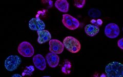
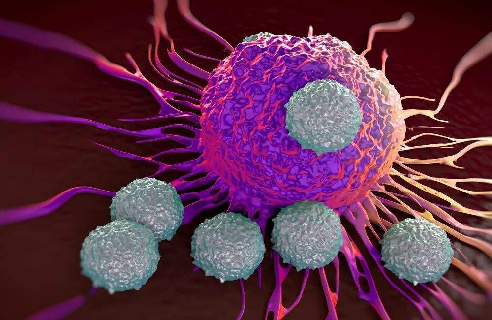
The body’s immune system eliminates tumor cells or inhibits their growth in a variety of ways. The body’s anti-tumor immune response includes cellular immunity and humoral immunity.
1. Cellular immunity: It is the main way of tumor immune response. As a specific immune response, it mainly produces an immune response to solid tumor cells with strong antigenicity.
2. Humoral immunity: play a synergistic role. As a non-specific immune response, circulating antibodies and the like mainly generate immune responses against tumor cells with weak antigenicity and free state.
tumor immune escape
Although there are a series of immune surveillance mechanisms in the body, it is still difficult to prevent the occurrence and development of tumors. A small number of tumor cells are not easy to cause the body’s response, and when the tumor grows to a certain extent, beyond the ability of the body’s immune response, the tumor cells can escape. The mechanism of tumor immune escape involves multiple immune response links.
1. Immune selection: Tumor cells with strong antigenicity can cause an effective immune response to be eliminated, while tumor cells with relatively weak antigenicity can escape the surveillance of the immune system and proliferate.
2. Antigen modulation: The antigenicity on the surface of tumor cells is reduced or lost to evade the recognition and attack of the host immune system.
3. Tumors cause immunosuppression: when tumor cells first appear, the amount of antigens is small and weak, and continuous stimulation of the host can induce immune tolerance and make the host lose the ability to respond to the tumor.
4. Blocking factors: blocking factors that cause immune promotion include blocking antibodies, excess free antigens and antigen-antibody complexes. The body can produce cytotoxic antibodies and blocking antibodies to tumors. Cytotoxic antibodies can cooperate with complement or NK cells to kill and lyse tumor cells. However, the binding of antigens shed by tumor cells to these antibodies prevents the cytotoxic effects of these antibodies. The binding of blocking antibodies to tumor cells is not only harmless to tumors, but also prevents the killing effect of sensitized lymphocytes and cytotoxic antibodies on tumor cells.
Tumor Immunodiagnosis
Tumor markers are detected by biochemical and immunological techniques to assist in the diagnosis of tumors. Common tumor markers include tumor antigens, hormones, oncogenes and their expressed proteins.
1. Embryonic antigens
Embryonic antigens are synthesized by embryonic tissues and exist in fetal serum and amniotic fluid. They will decrease after birth, but they will be greatly increased when certain tumors occur, which are called embryonic antigens. Embryonic tumor markers include alpha-fetoprotein (AFP) and carcinoembryonic antigen (CEA).
(1) Alpha-fetoprotein (AFP) is synthesized by hepatocytes and yolk sac during the embryonic period, and exists in fetal serum. The fetal serum content is the highest in 4-5 months, and then gradually decreases with the increase of gestational age, and decreases rapidly after birth. disappear. The content of normal adult serum is extremely low, and the normal reference value of AFP is <25ng/ml.
Elevated AFP levels are commonly seen in: ① Primary liver cancer. When primary liver cancer occurs, about 70% of patients have elevated AFP levels in serum, but some patients have normal AFP levels; ② Liver cirrhosis and viral hepatitis, patients with serum AFP levels The content of AFP will increase in different degrees; (3) Pregnancy, after 3 months of pregnancy, the level of serum AFP increases, peaks at 7 to 8 months, and returns to normal 3 weeks after delivery; (4) Reproductive system tumors and embryonic tumors , teratoma, testicular cancer patients with AFP levels are often elevated.
(2) Carcinoembryonic antigen (CEA) exists in the gastrointestinal tract, pancreas and liver of fetuses from 2 to 6 months old, and the content in postnatal tissues is very low, with a normal reference value of <5.0ng/ml.
Elevated CEA is common in patients with gastrointestinal (colon, rectum, pancreas) malignant tumors, breast cancer, lung cancer and other malignant tumors.
2. Serum Ferritin (SF)
Diagnosis of liver, lung, pancreas and other cancers, acute leukemia, etc.
3. Carbohydrate antigen series (CA) recognized by monoclonal antibodies
Sugar chain antigens are tumor-related antigens recognized by monoclonal antibodies prepared from various tumor cell lines. Most of them are glycoproteins, and they mostly exist on the surface of tumor cells. Such as CA50, CA125, CA15-3, CA19-9, CA72-4, CA130, CA242 and so on.
(1) CA50 is a glycolipid antigen mainly composed of sialic acid lipids and sialic acid glycoproteins, which plays an important role in the information transmission, growth and differentiation of normal cells. It is mainly used for auxiliary diagnosis and progress monitoring of pancreatic cancer, colorectal cancer, gastric cancer, liver cancer, etc.
(2) CAl25 is a glycoprotein that exists in epithelial ovarian cancer tissues and serum of patients. It is mainly used to assist in the diagnosis of ovarian cancer, but it is found in ovarian cysts, endometriosis, lung cancer, benign and malignant pleural ascites. Positive reactions were also seen.
(3) CAl5-3 is a breast cancer-related antigen, which has a certain value in the diagnosis of breast cancer and postoperative follow-up monitoring, but its sensitivity is low in the early stage of breast cancer.
(4) CAl9-9 exists in fetal pancreas, gallbladder, liver, intestine and other tissues, and the content in normal human tissues is extremely low. The content of CAl9-9 in the serum of patients with malignant tumors of the digestive tract is significantly increased, and the detection of serum CAl9-9 can be used as an auxiliary diagnostic index for malignant tumors such as gastric cancer, pancreatic cancer, and gallbladder cancer.
(5) CA72-4 is a macromolecular glycoprotein carcinoid antigen, which is a marker for gastrointestinal tumors and ovarian cancer. It can be used to diagnose gastric cancer, ovarian cancer, colorectal cancer, breast cancer, pancreatic cancer, etc.
(6) CA130 can be used to diagnose ovarian cancer, endometrial cancer, etc.
(7) CA242 can be used to diagnose gastrointestinal tumors, etc.
4. Hormones
When tissues that do not produce hormones undergo malignant transformation, they will produce and release some peptide hormones.
(1) Human chorionic gonadotropin (HCG) is a glycoprotein hormone secreted by placental trophoblast cells, and complete HCG is produced by the syncytiotrophoblast of placental chorion. HCG detection is an important indicator for monitoring early pregnancy. Under abnormal conditions, trophoblastic tumors and germ cell tumors, such as moles and malignant moles, choriocarcinoma and testicular teratocarcinoma, can significantly increase HCG.
(2) Calcitonin (CT) is mainly a polypeptide hormone secreted by thyroid follicular C cells. The main function of calcitonin is to reduce blood calcium content. Elevated levels are common in medullary thyroid cancer, small cell lung cancer, pancreatic cancer, uterine cancer, breast cancer, prostate cancer, benign thyroid cell adenomas, and acute or chronic renal failure. Reduction is common in severe hyperthyroidism and thyroid hypoplasia.
5. Enzymes
Including prostate-specific antigen, prostatic acid phosphatase, neuron-specific enolase and α-L-fucosidase and so on. Enzymes are one of the earlier tumor markers used for clinical diagnosis.
(1) The normal value of prostate-specific antigen (PSA) is less than 4 μg/L, and the positive rate in prostate cancer is as high as 30% to 86%, and its elevated level is closely related to the tumor.
(2) The normal value of neuron-specific enolase (NSE) is less than 15 μg/L. It is currently considered a tumor marker for small cell lung cancer and neuroblastoma. Elevated levels of NSE in serum are commonly seen in: small cell lung cancer, neuroblastoma, neuroendocrine cell tumors, and hypoxic-ischemic brain injury.
(3) α-L-fucosidase (AFU) 234-414 μmol/L. It is mainly used for the diagnosis of hereditary AFU deficiency and to differentiate it from other hereditary mucopolysaccharidosis diseases. Serum AFU was significantly increased in patients with primary liver cancer, chronic hepatitis, liver cirrhosis, bile duct cancer, colon cancer, uterine cancer, breast cancer, and lung cancer. Serum AFU also increases in women during pregnancy, but decreases rapidly after delivery.
tumor immunotherapy
1. Active Immunotherapy
Therapeutic approaches to stimulate and enhance host antitumor immune responses. Active immunotherapy is divided into specific and non-specific categories, the former uses tumor-specific antigens, and the latter uses substances that stimulate the immune system non-specifically. Active immunotherapy is suitable for immune-responsive hosts and/or immunogenic tumors.
2. Passive Immunotherapy
A therapy for treating tumors by infusing the host with conjugates of effector cells and/or antibodies that can directly kill tumors. When the therapeutic factor is cells, it is called adoptive immunotherapy. Passive immunotherapy does not depend on the immune functional status of the host.