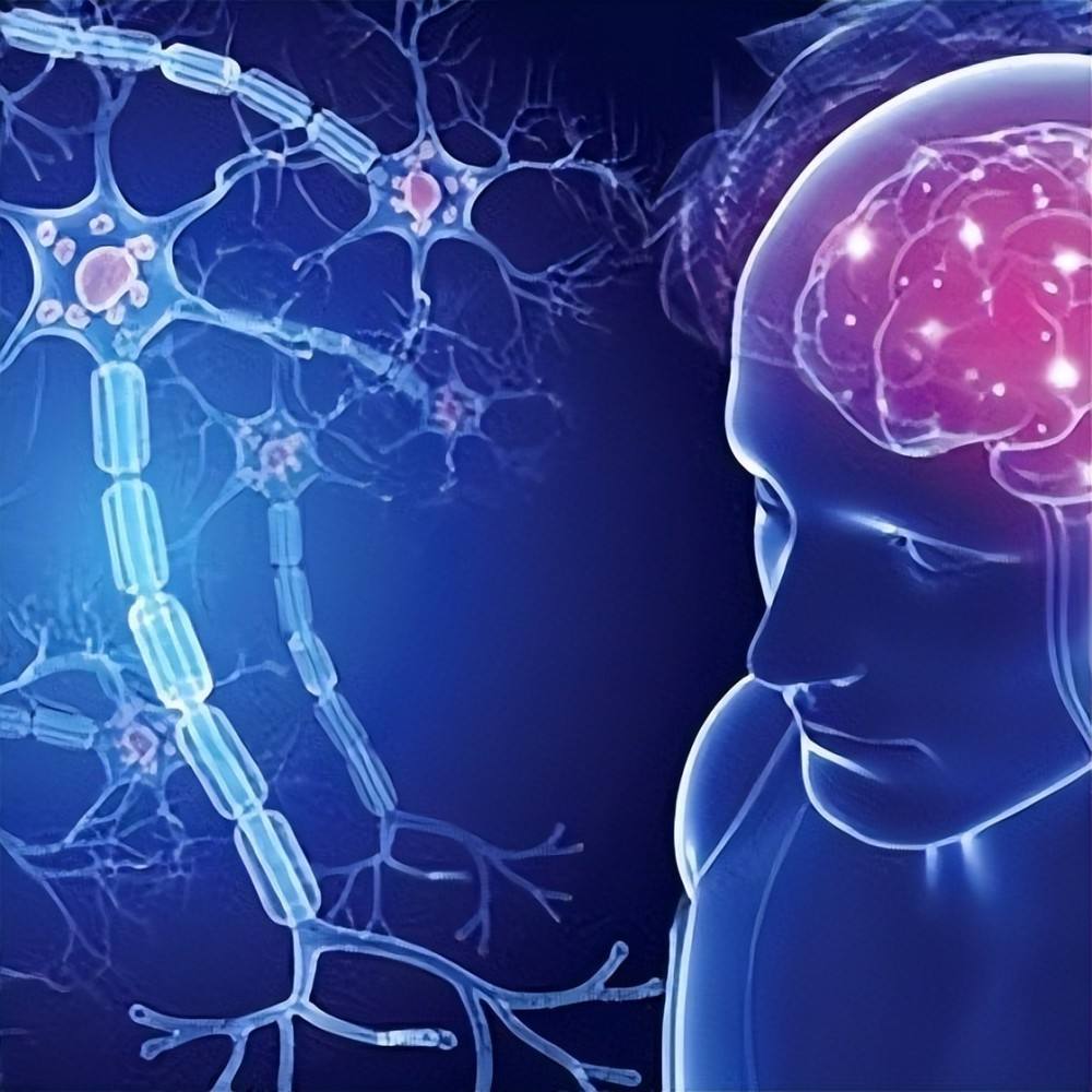
Multiple sclerosis is an autoimmune disease that causes damage to axons and neurons throughout the nervous system. While this damage is often unnoticed by patients at first, the extent of the damage determines the prognosis for disease severity. Predictive judgment of the disease process is important for selecting appropriate multiple sclerosis treatments, and medical research is constantly searching for reliable prognostic tools.
Recently, a research team from the Medical University of Vienna and the University of Vienna has shown for the first time that retinal layer thinning can be used as a prognostic marker.
In the new study, the researchers studied 167 MS patients with an average age of 36.5 years (average duration of disease 3.1 years) over a three-year period. It was observed that these patients all experienced an acute optic neuritis episode and at least one subsequent non-optic neuritis recurrence. The researchers measured the retinal layer thicknesses of the patients using optical coherence tomography (OCT).
They speculate that retinal thinning caused by multiple sclerosis relapses could reflect the extent of brain damage. The analysis confirmed that a loss of retinal layer thickness of approximately 5 µm (micrometers) after optic neuritis corresponds to a doubling of the risk of permanent disability after the next recurrence. Therefore, retinal layer thinning can be used as a biomarker of neurological damage in multiple sclerosis, measuring retinal layer thickness at the initial diagnosis of multiple sclerosis and again six months later, through layer difference can predict disability degree of deterioration.
The researchers look forward to the study showing that thinning of the retinal layer after optic neuritis may be a marker of future relapsing remission in relapsing forms of multiple sclerosis. This study highlights the value of retinal layer thinning in prognostic applications in multiple sclerosis. If this result is confirmed in a larger follow-up study, the technique could be applied in clinical practice.