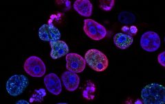
Cytotoxicity is a purely cell-killing event caused by cells or chemicals that does not rely on apoptosis or necrosis as a cell death mechanism. Testing for the cytotoxicity of specific substances, such as drug screening, is sometimes required.
Cytotoxicity detection is mainly based on changes in cell membrane permeability. The following methods are commonly used:
MTT and XTT methods: using the activity of enzymes inside the mitochondria, specific tetrazolium salts can be converted, and then detected by a microplate reader;
LDH method: cytotoxicity is detected by detecting the enzymatic activity of LDH in the cell culture supernatant;
Other enzymatic methods: such as detecting the activities of alkaline phosphatase and acid phosphatase in the supernatant;
Cell Proliferation Assay Kit:
Principle: Normal cells have vigorous metabolism, and the succinate dehydrogenase in their mitochondria can reduce tetrazolium salts (such as MTT, XTT, WST-1, etc.) to purple crystalline substances, which are deposited around the cells, and then Read the OD value by a microplate reader to detect the cell proliferation state
Fluorescein luminescence assay for cell viability
Principle: Adenylate kinase (AK) exists in the cytoplasm of all eukaryotic and prokaryotic cells, and AK has the ability to activate ADP to generate ATP. When cells are damaged, the cell membrane is damaged and AK is released into the culture supernatant. The kit utilizes luciferase and luciferin to emit light under the action of ATP, and can be quantitatively detected by a chemiluminescence instrument.
Cytotoxicity assay by LDH method
Principle: LDH (lactate dehydrogenase) is a stable protein that exists in the cytoplasm of normal cells. Once the cell membrane is damaged, LDH is released outside the cell; LDH catalyzes the formation of pyruvate from lactate, and INT (four azole salts) to form a purple crystalline substance, which can be detected by a 500nm microplate reader. By detecting the activity of LDH in the cell culture supernatant, the degree of cell damage can be judged.
explain
“Cytotoxicity” Explained in Academic Literature
1. According to the definition of document CEN1992 No. 30 of the European Organization for Standardization, cytotoxicity refers to cell death, cell lysis and cell growth inhibition caused by products, materials and their impregnants.
2. Cytotoxicity refers to the toxicity of the substance to cultured cells. P-388LulZ0 leukemia cells or KB tumor cells are usually used for in vitro tests. To test the cytotoxicity of marine natural products, sea urchin fertilized eggs are often used to observe the inhibitory effect of the samples on their cell division.
3. It has strong cytotoxicity and promotes the proliferation of these two kinds of cells, also known as cytotoxicity
Classification and application
Cytotoxicity is the effect of chemical substances (drugs) on the basic structure and/or physiological process of cells, such as cell membrane or cytoskeleton structure, cell metabolism process, synthesis, degradation or release of cell components or products, ion regulation and cell division and other processes , leading to disorders of cell survival, proliferation and/or function, resulting in adverse reactions. According to the mechanism of action, it can be divided into 3 types:
①Basic cytotoxicity, involving one or more of the above structural or functional changes, acting on all types of cells;
②Selective cytotoxicity, which exists on some differentiated cells, is mainly caused by the biotransformation of chemical substances, binding to special receptors or a special intake mechanism;
③ Special cell function toxicity, slight damage to cell structure and function, but very serious damage to the whole body. Similar toxic effects can be achieved through the synthesis, release, binding and degradation of cytokines, hormones and transmitters affecting cell-to-cell communication or special transport processes. Toxic effects may also arise from chemical interference with extracellular processes, and any non-animal assay system has multiple factors to consider. In 1983, Ekwall put forward the concept of “basic cell function”, that is, the toxic effects of most chemical substances are non-specific damage to cell function, but can cause specific changes in organ function and even death of the body.
Studies have shown that there is a good correlation between the in vitro cytotoxicity of chemical substances and their plasma concentrations of animal mortality and human death. Chemical damage and death can ultimately manifest as changes at the cellular level, suggesting that in vitro cytotoxicity predicts acute in vivo toxicity. More than 50 years from earlier studies, in vitro systems that can predict acute toxicity in vivo have been developed. The quantitative study between in vitro cytotoxicity and acute toxicity is mainly based on the in vitro cytotoxicity IC50 value and in vivo acute toxicity of various chemicals in the RC (Registry of Cytotoxicity) database (National Institute of Occupational Safety and Health Chemical Substances Toxicity Database). Toxicity LD50 value was correlated and analyzed, and RC prediction model was obtained for prediction of acute toxicity LD50 value. Using cytotoxicity to compare the predictive ability of various detection endpoints, different tissues and species, it was found that rodent cell lines have good predictive ability for acute toxicity in rodents and human cell lines for human acute toxicity. BALB/c3T3 cells and human normal keratinocytes (NHK) have good stability and predictive ability in validation experiments, so they are recommended as common cell lines for cytotoxicity analysis, and other cell lines and detection endpoints can also be used. In vitro methods help predict systemic and local effects from acute exposure to chemicals and assess toxic concentrations in vivo. Therefore, in vitro cytotoxicity analysis was performed before the acute toxicity test, and then the LD50 value was predicted according to the RC prediction model, and the most suitable starting dose for acute toxicity in vivo was selected to reduce the use of experimental animals.