Cancer is unpredictable. The tumor shrinks after the initial treatment, but it can become more terrifying afterward. The disease can spread or mutate, and genetic changes make the cancer more sensitive to treatment options, or vice versa.
Oncologists often rely on invasive biopsies and non-invasive imaging modalities to track tumor size, expansion, and response to therapy. However, circulating tumor cells (CTCs) offer a promising new approach. Here, we review methods to isolate and study these tumor vanes.
CTC Definition
Circulating tumor cells are cancer cells that shed from a tumor and enter the circulatory system, where they can be found with a simple blood test. These cells serve as biomarkers for cancer, indicating the presence or absence of distal tumors, as well as their genetic status and drug susceptibility. Unlike surgical intervention, CTC is compatible with longitudinal studies and can effectively guide treatment. However, to understand their meaning, it is not so simple.
CTCs, like the tumors themselves, are highly heterogeneous, says Zena Werb, a professor of anatomy at the University of California, San Francisco. Some CTCs are capable of generating new tumors, while others are not. Some exhibited properties of cancer stem cells, while others did not. Currently, the only clinical test for CTCs approved by the US FDA is Janssen Diagnostics’ CellSearch, which effectively defines epithelial-derived CTCs that are positive for EpCAM, DAPI, and cytokeratin, but negative for CD45.
Of course, not all tumors originate from epithelial cells, and even epithelial tumors may gradually lose epithelial markers (CellSearch is approved for metastatic breast, colorectal, and prostate cancer). And there are indications that, in addition to cancer, inflammation may also lead to the presence of epithelial cells in the blood. Werb believes the story is more complicated than we thought.
According to Ron Mazumder, head of research and development at Janssen Diagnostics, the proportion of patients with detectable CTCs in their blood varies by cancer type. For example, colorectal, ovarian and breast cancer is about 50-70%, while non-small cell lung cancer is as low as 30%. The numbers of CTCs in these patients also varied widely, from a single cell to several thousand. “The median is about 20 to 30 cells,” Mazumder said, but they found that metastatic breast and prostate cancer patients with five or more CTCs per 7.5 ml of blood had a worse prognosis than CTCs. Fewer patients are much worse. Therefore, five was set as the cutoff value – if the number of CTCs was greater or less, this had a significant impact on patient survival, and this was true across multiple cancer types.
Microfluidic options
Originally, CellSearch relied entirely on cell counting. Today, users also have access to the genetic information of these cells. In August 2014, Janssen announced a partnership with Asuragen to perform next-generation sequencing of isolated CTCs to identify mutations in hundreds of cancer-related genes.
CellSearch is based on positive selection, using antibody-coated magnetic beads to capture EpCAM-positive cells, which are then imaged and counted. So, says Shannon Stott, an assistant professor at the Massachusetts General Hospital Cancer Research Center, you need to know in advance the properties of the cells you’re looking for. However, not all CTCs have the same physical and biological properties.
Stott has a background in mechanical engineering, and in collaboration with several colleagues, she developed new methods for isolating CTCs. In 2010, she developed a “herringbone CTC chip” that uses EpCAM antibodies to capture CTCs. The design, she says, can process blood relatively quickly (2 ml per hour) with good sensitivity and selectivity. However, it cannot account for the different antigenic profiles. The captured cells stick to the surface of the chip, making subsequent culturing problematic.
In 2013, a team at Massachusetts General Hospital developed a third-generation technology called CTC¬iChip. The i in this iChip stands for inertial focusing, where the cells are aligned in a column by fluid forces. The chip utilizes hydrodynamic sorting to remove the smallest blood components. Then, it uses positive selection to remove lymphocytes and keep everything that is left.
According to Stott, the iChip is much faster than the chevron chip, processing 20 ml of blood per hour. It also yields unlabeled cells for easy culture and analysis. Her colleague Daniel Haber’s team established CTC cell lines from six breast cancer patients. Sequencing of these cell lines identified newly acquired mutations and drug susceptibility, information that could help develop better therapeutic strategies. In another study, Haber’s team performed single-cell RNA¬seq on isolated CTCs and compared them to CTC aggregates from the same patient to understand the nature of these clustered CTC structures. Clustered CTCs have greater metastatic potential than individual CTCs. The analysis identified a cell junction-related gene called plakoglobin.
Currently, Janssen Diagnostics is working with a team at Massachusetts General Hospital to transform the iChip into a next-generation CTC analysis platform.
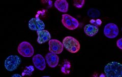
CTC imaging
An article in Nature Reviews Clinical Oncology reviews the principles of CTC analysis and identification. There is a table in the article that lists selected detection techniques, most of which are based on the antigenic profile or physical properties of CTCs. An alternative strategy mentioned in the table is image flow cytometry, which is commercialized by Merck Millipore.
Both ImageStream and FlowSight are image flow cytometers, which are essentially flow cytometers with cameras. It images in brightfield and darkfield modes and up to 10 channels of fluorescence as each cell passes through, producing 12 images of each cell and capable of imaging 2,000 cells per second. The difference between the two platforms is that ImageStream can zoom up to 60x, while FlowSight offers a fixed 20x magnification.
Although the resulting cells cannot be cultured, users can perform various analyses as the cells pass through the system. For example, they can stain for extracellular and intracellular markers to identify cells of origin, or stain with RNA-FISH probes to count transcripts.
Of course, traditional flow cytometry can also obtain many parameters, and image flow cytometry can give you confidence that these few positive cells are CTCs. “Seeing is believing,” said David Basiji, senior director of Millipore Cell Analysis at Merck. Today, the company is working to isolate CTCs for downstream research.
References:
Stott, SL, et al., “Isolation of circulating tumor cells using a microvortex¬generating herringbone¬chip,” Proc Natl Acad Sci USA, 107:18392–7, 2010. [PubMed: 20930119]
Ozkumur, E, et al., “Inertial focusing for tumor antigen¬dependent and –independent sorting of rare circulating tumor cells,” Sci Transl Med, 5:179ra47, 2013. [PubMed: 23552373]
Yu, M, et al., “Ex vivo culture of circulating breast tumor cells for individualized testing of drug susceptibility,” Science, 345:216–220, 2014. [PubMed: 25013076]
Aceto, N, et al., “Circulating tumor cell clusters are oligoclonal precursors of breast cancer metastasis,” Cell, 158:1110–22, 2014. [PubMed: 25171411]
Krebs, MG, et al., “Molecular analysis of circulating tumour cells—biology and biomarkers,” Nat Rev Clin Oncol, 11:129–44, 2014. [PubMed: 24445517]